
Image Type
Segmental Angles
1) Description of Measurement
Angulation between segments quantifies the relative sagittal plane rotation between two adjacent cervical vertebrae and is used to detect segmental instability, fracture–dislocation patterns, and ligamentous disruption.
This measurement reflects abnormal flexion or extension deformity at a motion segment and is a core criterion in the radiographic definition of cervical spinal instability, particularly in trauma settings.
2) Instructions to Measure
Obtain thin-slice cervical CT with high-quality sagittal reconstructions.
Identify the motion segment of interest (e.g., C4–C5).
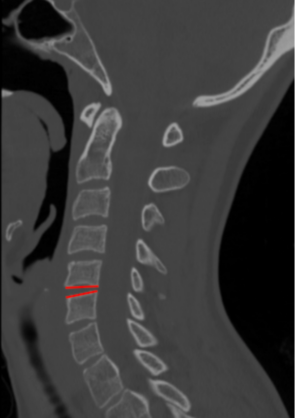
Draw a line along the inferior endplate of the superior vertebra (e.g., inferior endplate of C4).
Draw a second line along the superior endplate of the inferior vertebra (e.g., superior endplate of C5).
Repeat at suspicious levels and record the largest angulation.
3) Normal vs. Pathologic Ranges
Normal/physiologic motion: ≤ 11°
Pathologic angulation: > 11°; indicates instability
Key points:
> 11° of angulation at a single motion segment is a classic radiographic criterion for cervical instability
Greater angulation correlates with ligamentous disruption and higher risk of neurologic compromise
4) Important References
Bono CM, Vaccaro AR, Fehlings M, Fisher C, Dvorak M, Ludwig S, Harrop J; Spine Trauma Study Group. Measurement techniques for upper cervical spine injuries: consensus statement of the Spine Trauma Study Group. Spine (Phila Pa 1976). 2007 Mar 1;32(5):593-600. doi: 10.1097/01.brs.0000257345.21075.a7.
White AA, Panjabi MM. Clinical Biomechanics of the Spine. 2nd ed. Lippincott; 1990.
Harris JH Jr, Mirvis SE. The Radiology of Acute Cervical Spine Trauma. 3rd ed. Williams & Wilkins; 1996.
Dvorak MF, Fisher CG, Fehlings MG, Rampersaud YR, Oner FC, Aarabi B, Vaccaro AR. The surgical approach to subaxial cervical spine injuries: an evidence-based algorithm based on the SLIC classification system. Spine (Phila Pa 1976). 2007 Nov 1;32(23):2620-9. doi: 10.1097/BRS.0b013e318158ce16.
Daffner RH, Sciulli RL, Rodriguez A, Protetch J. Imaging for evaluation of suspected cervical spine trauma: a 2-year analysis. Injury. 2006 Jul;37(7):652-8. doi: 10.1016/j.injury.2006.01.018.
5) Other info....
CT is excellent for detecting bony malalignment; MRI should be obtained when abnormal angulation is present to evaluate:
Disc disruption
Posterior ligamentous complex injury
Spinal cord compression
Even angulation just above the 11° threshold may be clinically significant in the presence of neurologic symptoms or additional instability markers
In multilevel injuries, the most angulated segment dictates instability grading and management strategy
Adapted from: Thibodeau R, Normal CT cervical spine. Case study, Radiopaedia.org (Accessed on 28 Dec 2025) https://doi.org/10.53347/rID-183532