Image Type
Spinolaminar Line
1) Description of Measurement
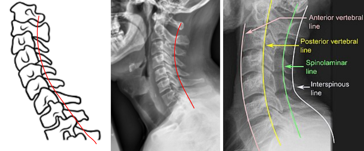
The Spinolaminar Line is a radiographic reference line used to assess posterior cervical alignment and detect subluxation, dislocation, or instability.
It is drawn along the junction between the laminae and spinous processes (the spinolaminar junction) of consecutive cervical vertebrae on a lateral cervical spine X-ray.
A smooth, continuous curve from C1 to C7 represents normal posterior alignment. Any step-off or discontinuity indicates possible vertebral displacement, facet dislocation, or fracture, particularly in traumatic injuries.
This line complements George’s Line (posterior vertebral body line) as part of a systematic three-line evaluation for cervical stability.
2) Instructions to Measure
Obtain a neutral lateral cervical spine X-ray (as shown in the attached image).
Identify the spinolaminar junction points for each vertebra (C1–C7).
The spinolaminar junction lies where the lamina meets the base of the spinous process, appearing as a continuous posterior cortical shadow.
Draw a smooth, curving line connecting these junction points from C1 through C7.
Evaluate the line for continuity and symmetry:
A continuous curve indicates normal posterior alignment.
A posterior step-off (>1–2 mm) between adjacent vertebrae suggests posterior displacement or subluxation.
Repeat assessment on flexion and extension radiographs to evaluate for dynamic instability if indicated.
3) Normal vs. Pathologic Ranges
Normal alignment: smooth, uninterrupted curve; intact posterior cervical alignment
Normal variation: step-off ≤ 1mm; mild positional difference, no instability
Abnormal alignment: step-off > 1-2 mm; suggestive of subluxation or malalignment
Pathologic alignment: step off > 3.5 mm; diagnostic for cervical instability or fracture-dislocation
4) Important References
White AA, Panjabi MM. Clinical Biomechanics of the Spine. 2nd ed. Philadelphia: Lippincott; 1990.
Harris JH Jr, Edeiken-Monroe B. The Radiology of Emergency Medicine. 3rd ed. Baltimore: Williams & Wilkins; 1993.
Rogers LF. Radiology of Skeletal Trauma. 4th ed. Philadelphia: Saunders; 2014.
Lee C, Woodring JH, Rogers LF, Kim KS. Sagittal alignment of the cervical spine: significance of the posterior vertebral and spinolaminar lines. AJR Am J Roentgenol. 1986;146(3):707–714.
Scher AT. Displacement of the spinolaminar line–a sign of value in fractures of the upper cervical spine. S Afr Med J. 1979 Jul 14;56(2):58-61.
5) Other info....
The Spinolaminar Line, along with George’s Line (posterior vertebral line) and the anterior vertebral body line, forms the three-line cervical alignment assessment on lateral X-ray.
A disruption in the spinolaminar line can indicate:
Unilateral or bilateral facet dislocation
Ligamentous injury (posterior ligamentous complex disruption)
Spondylolisthesis or fracture malalignment
The line is particularly useful in trauma settings to detect subtle malalignment not obvious on the vertebral body lines alone.
CT or MRI should be obtained for confirmation if a break is observed.
Always correlate findings with clinical presentation and neurological status.