
Image Type
Power’s Ratio
1) Description of Measurement
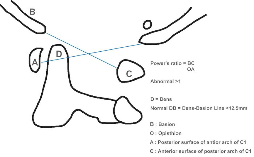
Power’s Ratio is a radiographic measurement used to assess atlanto-occipital dissociation (AOD), an unstable and potentially fatal injury involving separation between the skull and the cervical spine.
It evaluates the relationship between the skull base and the atlas (C1) by comparing the anterior and posterior distances between the foramen magnum (basion and opisthion) and the atlas (anterior arch and spinolaminar line).
A normal ratio suggests intact ligamentous connections between the occiput and atlas; an increased ratio indicates anterior displacement of the skull relative to the cervical spine, characteristic of AOD.
2) Instructions to Measure
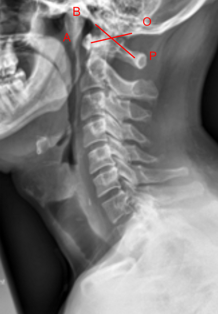
Obtain a neutral lateral cervical spine X-ray (as shown in the provided image).
Identify the following landmarks:
Basion (B): The anterior margin of the foramen magnum.
Opisthion (O): The posterior margin of the foramen magnum.
Anterior arch of the atlas (A): The midpoint of its anterior cortex.
Posterior arch of the atlas/spinolaminar line (P): The midpoint of its posterior cortex.
Draw two straight lines:
Line 1 (BA): From the basion (B) to the midpoint of the anterior arch of C1 (A).
Line 2 (OP): From the opisthion (O) to the midpoint of the posterior arch of C1 (P).
Measure the lengths of BA and OP.
Calculate Power’s Ratio = BA / OP.
BA = anterior basion-to-atlas distance.
OP = posterior opisthion-to-atlas distance.
3) Normal vs. Pathologic Ranges
Power’s Ratio ≤ 1.0: Normal occipitoatlantal alignment
Power’s Ratio > 1.0: Abnormal; suggests anterior displacement of the skull on the atlas (possible AOD)
Power’s Ratio > 1.2: Highly suggestive of atlanto-occipital dislocation; usually indicates disruption of the tectorial membrane, alar ligaments, or cruciate complex
4) Important References
Powers B, Miller MD, Kramer RS, Martinez S, Gehweiler JA Jr. Traumatic anterior atlanto-occipital dislocation. Neurosurgery. 1979;4(1):12–17.
Traynelis VC, Marano GD, Dunker RO, Kaufman HH. Traumatic atlanto-occipital dislocation: case report and review of the literature. Neurosurgery. 1986;19(1):112–117.
Harris JH Jr, Carson GC, Wagner LK, Kerr N. Radiologic diagnosis of traumatic occipitovertebral dissociation: 1. Normal occipitovertebral relationships on lateral radiographs of adults. AJR Am J Roentgenol. 1994;162(4):881–886.
Smoker WRK. Craniovertebral junction: normal anatomy, craniometry, and congenital anomalies. Radiographics. 1994;14(2):255–277.
5) Other info....
Power’s Ratio is simple and reproducible, making it valuable in the initial trauma evaluation of patients with suspected upper cervical injury.
Because it depends on accurate landmark visualization, CT or MRI confirmation is recommended for definitive diagnosis.
Additional measurements such as the Basion-Dens Interval (BDI) and Basion-Axial Interval (BAI) should be obtained concurrently for comprehensive assessment.
In pediatric patients, normal ligamentous laxity can lead to ratios slightly above 1.0 — interpret cautiously.
Always assess in the context of clinical stability and neurological findings.
In some posteriorly displaced injuries, the ratio may be < 0.7, indicating posterior translation of the skull relative to the atlas.
Richards PJ. Cervical spine clearance: a review. Injury. 2005 Feb 1;36(2):248-69.