
Image Type
Facet Joint Overlap (Percent Overlap Method)
1) Description of Measurement
Facet joint overlap (percent overlap method) is a quantitative CT-based assessment of cervical facet articulation used to identify facet fractures, subluxation, dislocation, and instability, particularly in the trauma setting.
The method expresses how much of the inferior articular facet of the superior vertebra remains overlapped with the superior articular facet of the inferior vertebra, normalized to the full anteroposterior length of the facet. Loss of overlap reflects posterior element disruption and correlates with mechanical instability and risk of neurologic injury.
2) Instructions to Measure
Obtain thin-slice cervical CT with sagittal and/or oblique reconstructions optimized to visualize the facet joints.
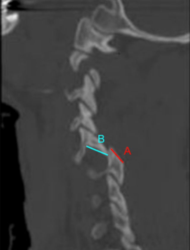
Select the facet level of interest, ideally the level with suspected injury or maximal malalignment.
Identify the articulating facets:
Inferior articular facet of the superior vertebra
Superior articular facet of the inferior vertebra
Measure Facet Length (A):
Measure the total anteroposterior length of the superior articular facet of the inferior vertebra.
Measure Overlap Length (B):
Measure the portion of the inferior articular facet that remains overlapped with the superior articular facet in the anteroposterior direction.
Calculate Percent Facet Overlap:
Facet Overlap (%) = (BA)100
Perform measurements bilaterally; record the lowest overlap percentage, as this represents the worst instability.
3) Normal vs. Pathologic Ranges
Normal facet articulation: ≥ 50%
Partial subluxation: 25-49%; potentially unstable
Severe instability: <25%
Facet dislocation: 0%; perched or jumped facet
Key points:
< 50% overlap is generally considered abnormal
Unilateral loss may indicate rotational injury
Bilateral loss strongly suggests gross instability
4) Important References
Dvorak MF, Fisher CG, Fehlings MG, et al. The surgical approach to subaxial cervical spine injuries: an evidence-based algorithm based on the SLIC classification system. Spine (Phila Pa 1976). 2007 Nov 1;32(23):2620-9. doi: 10.1097/BRS.0b013e318158ce16.
Daffner RH, Sciulli RL, Rodriguez A, Protetch J. Imaging for evaluation of suspected cervical spine trauma: a 2-year analysis. Injury. 2006 Jul;37(7):652-8. doi: 10.1016/j.injury.2006.01.018.
Allen BL Jr, Ferguson RL, Lehmann TR, O'Brien RP. A mechanistic classification of closed, indirect fractures and dislocations of the lower cervical spine. Spine (Phila Pa 1976). 1982 Jan-Feb;7(1):1-27. doi: 10.1097/00007632-198200710-00001.
Harris JH Jr, Mirvis SE. The Radiology of Acute Cervical Spine Trauma. 3rd ed. Williams & Wilkins; 1996.
5) Other info....
Facet overlap is a direct structural measure of stability, unlike purely alignment-based metrics
Particularly useful for:
Facet fractures
Perched or jumped facets
Rotational or distraction injuries
Should be interpreted alongside:
Anterior/posterior translation
Interspinous widening
Disc space disruption
Neurologic findings
CT is the gold standard for bony facet evaluation; MRI complements this by assessing ligamentous and disc injury
Even with preserved overlap, associated ligamentous disruption may still confer instability—clinical correlation is essential
Adapted from: Foresthoefel C, Moore WD. Cervical Facet Dislocations and Fractures. Reference article, Orthobullets. (Accessed on December 20, 2025). https://www.orthobullets.com/spine/2064/cervical-facet-dislocations-and-fractures