
Image Type
Sagittal Vertical Axis (SVA)
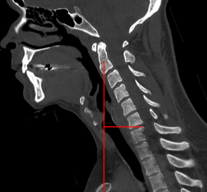
1) Description of Measurement
Cervical sagittal vertical axis (SVA) quantifies the anterior–posterior global alignment of the cervical spine by measuring the horizontal offset between a cranial reference point and a caudal cervical landmark. It reflects the forward or backward translation of the head over the cervical spine and is a key parameter in the evaluation of cervical sagittal balance, deformity, and postoperative outcomes.
Increased cervical SVA correlates with neck pain, disability, impaired horizontal gaze, and poor health-related quality of life.
2) Instructions to Measure
Obtain a true mid-sagittal CT reconstruction showing the skull base through C7.
Identify the center of the C2 vertebral body.
From this point, drop a vertical plumb line parallel to the vertical axis of the image.
Identify the posterior–superior corner of the C7 vertebral body.
Measure the horizontal distance (mm) between the C2 plumb line and the posterior–superior corner of C7.
Record this value as the C2–C7 SVA.
3) Normal vs. Pathologic Ranges
Normal sagittal alignment: ≤ 20 mm
Mild anterior malalignment: 21 - 40 mm
Severe sagittal imbalance: > 40 mm
Key points:
Increasing SVA correlates with worse Neck Disability Index (NDI) scores.
Postoperative correction toward normal values is associated with improved functional outcomes.
4) Important References
Tang JA, Scheer JK, Smith JS, et al; ISSG. The impact of standing regional cervical sagittal alignment on outcomes in posterior cervical fusion surgery. Neurosurgery. 2015 Mar;76 Suppl 1:S14-21; discussion S21. doi: 10.1227/01.neu.0000462074.66077.2b.
Ames CP, Blondel B, Scheer JK, et al. Cervical radiographical alignment: comprehensive assessment techniques and potential importance in cervical myelopathy. Spine (Phila Pa 1976). 2013 Oct 15;38(22 Suppl 1):S149-60. doi: 10.1097/BRS.0b013e3182a7f449.
Fujiwara H, Oda T, Makino T, et al. Impact of Cervical Sagittal Alignment on Axial Neck Pain and Health-related Quality of Life After Cervical Laminoplasty in Patients With Cervical Spondylotic Myelopathy or Ossification of the Posterior Longitudinal Ligament: A Prospective Comparative Study. Clin Spine Surg. 2018 May;31(4):E245-E251. doi: 10.1097/BSD.0000000000000619.
5) Other info....
Although traditionally measured on standing lateral radiographs, cervical SVA can be reliably estimated on upright CT or neutral-position CT reconstructions.
Should be interpreted alongside:
C2–C7 lordosis
T1 slope
Chin–brow vertical angle (CBVA)
Excessive cervical SVA often represents compensatory mechanisms for thoracolumbar deformity.
When SVA is markedly abnormal, full-spine imaging is recommended to evaluate global sagittal balance.
Adapted from: Feger J, Yap J, Knipe H, et al. CT cervical spine (protocol). Reference article, Radiopaedia.org (Accessed on 28 Dec 2025) https://doi.org/10.53347/rID-89993