
Image Type
Translation on Sagittal Reconstruction
1) Description of Measurement
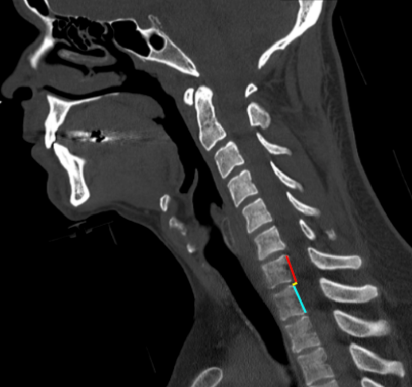
Translation on sagittal reconstruction quantifies the anteroposterior displacement of one cervical vertebral body relative to an adjacent vertebra on sagittal CT images. It is a direct indicator of spinal instability and reflects disruption of the anterior and/or posterior ligamentous complexes, often in the setting of trauma.
This measurement is particularly useful for identifying subluxation, fracture–dislocation, and unstable injury patterns, and it complements facet-based assessments (facet overlap, facet step-off) and global alignment lines.
2) Instructions to Measure
Obtain thin-slice cervical CT with high-quality sagittal reconstructions centered on the midline and parasagittal planes.
Identify the vertebral level of interest, particularly where malalignment is suspected.
On the sagittal image:
Identify the posterior cortical margin of the inferior vertebral body (reference vertebra).
Identify the posterior cortical margin of the superior vertebral body (translated vertebra).
Draw two vertical reference lines parallel to the sagittal axis:
One along the posterior margin of the inferior vertebral body.
One along the posterior margin of the superior vertebral body.
Measure the horizontal distance (mm) between these two lines.
This distance represents anteroposterior translation at that level.
Perform measurements at suspicious adjacent levels and record the greatest translation observed.
3) Normal vs. Pathologic Ranges
Normal or physiologic alignment: ≤ 2 mm
Borderline translation: 2 - 3.5 mm
Pathologic translation: > 3.5 mm; unstable injury
Key points:
> 3.5 mm of translation is a classic threshold for cervical instability
Larger degrees of translation correlate with disco-ligamentous disruption and increased neurologic risk
4) Important References
White AA, Panjabi MM. Clinical Biomechanics of the Spine. 2nd ed. Lippincott; 1990.
Harris JH Jr, Mirvis SE. The Radiology of Acute Cervical Spine Trauma. 3rd ed. Williams & Wilkins; 1996.
Dvorak MF, Fisher CG, Fehlings MG, Rampersaud YR, Oner FC, Aarabi B, Vaccaro AR. The surgical approach to subaxial cervical spine injuries: an evidence-based algorithm based on the SLIC classification system. Spine (Phila Pa 1976). 2007 Nov 1;32(23):2620-9. doi: 10.1097/BRS.0b013e318158ce16.
Daffner RH, Sciulli RL, Rodriguez A, Protetch J. Imaging for evaluation of suspected cervical spine trauma: a 2-year analysis. Injury. 2006 Jul;37(7):652-8. doi: 10.1016/j.injury.2006.01.018.
5) Other info....
Translation on sagittal CT is a direct, reproducible measure of instability
Particularly useful for detecting:
Fracture–dislocations
Facet dislocations
Ligamentous injuries not immediately obvious on axial images
CT is optimal for bony alignment; MRI is recommended when translation is present to assess:
Disc injury
Posterior ligamentous complex disruption
Spinal cord compression
Even translation just above the threshold may be clinically significant when associated with neurologic symptoms or additional instability markers
Adapted from: Feger J, Yap J, Knipe H, et al. CT cervical spine (protocol). Reference article, Radiopaedia.org (Accessed on 28 Dec 2025) https://doi.org/10.53347/rID-89993