
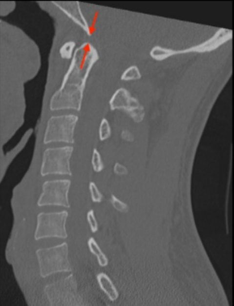
Image Type
Basion–Dens Interval (BDI)
1) Description of Measurement
BDI evaluates vertical alignment between the skull base and the upper cervical spine. It is primarily used to detect vertical distraction injuries and atlanto-occipital dissociation.
2) Instructions to Measure
Use a midsagittal CT image.
Identify the basion (anterior margin of the foramen magnum). Locate the lowest, most posterior point of the clivus in the midline.
Locate the most superior tip of the odontoid process.
Measure the distance from the basion to the tip of the dens.
In children under ~12 years, the tip of the dens may not be fully ossified (summit ossification center). In these cases, the ossified portion is used, but the metric loses reliability, often necessitating the use of the CCI.
3) Normal vs. Pathologic Ranges
The "Rule of 12s" (Radiography): Harris et al. established that on lateral cervical radiographs taken at a standard target-film distance (1 meter), the BDI should be < 12 mm in 95% of normal adults and children over 13. A value > 12 mm suggests AOD (specifically vertical distraction).
Plain radiographs involve geometric magnification (approx. 15-20%). MDCT images are reconstructed at 1:1 scale. Therefore, applying the 12 mm radiographic threshold to CT scans may yield false negatives.
CT Normal Values: Studies by Rojas et al. and others have defined the normal BDI on CT as <8.5-<10 mm. A BDI > 10 mm on CT is highly suspicious for AOD.
4) Important References
Bono CM, Vaccaro AR, Fehlings M, et al. Measurement techniques for upper cervical spine injuries: consensus statement of the spine trauma study group. Spine. 2007;32:593-600.
Daffner RH, Harris JH. Cervical Spine Injuries. 5th ed. Philadelphia, PA: Lippincott Williams & Wilkins; 2013:139–260
Harris JH Jr, Carson GC, Wagner LK. Radiologic diagnosis of traumatic occipitovertebral dissociation: 1. Normal occipitovertebral relationships on lateral radiographs of supine subjects. AJR Am J Roentgenol 1994;162(04):881–886
Harris JH Jr, Carson GC, Wagner LK, Kerr N. Radiologic diagnosis of traumatic occipitovertebral dissociation: 2. Comparison of three methods of detecting occipitovertebral relationships on lateral radiographs of supine subjects. AJR Am J Roentgenol 1994; 162(04):887–892
Joaquim AF, Schroeder GD, Vaccaro AR. Traumatic Atlanto-Occipital Dislocation-A Comprehensive Analysis of All Case Series Found in the Spinal Trauma Literature. Int J Spine Surg. 2021;15(4):724-739. doi:10.14444/8095
Rojas CA, Bertozzi JC, Martinez CR, Whitlow J. Reassessment of the craniocervical junction: normal values on CT. AJNR Am J Neuroradiol. 2007;28(9):1819-1823. doi:10.3174/ajnr.A0660
Adapted from: Knipe H, Atlantodental interval. Radiopaedia.org (Accessed on 17 Dec 2025). https://doi.org/10.53347/rID-38418