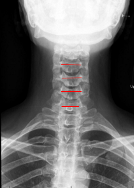
Image Type
Interpedicular Distance (IPD)
1) Description of Measurement
The Interpedicular Distance (IPD) is a radiographic measurement used to assess the transverse (mediolateral) width of the spinal canal at a given vertebral level. It represents the horizontal distance between the medial borders of the pedicles on an anteroposterior (AP) cervical spine X-ray.
This measurement is important for evaluating congenital, traumatic, or acquired narrowing of the spinal canal and is especially useful in detecting developmental cervical stenosis, fractures involving pedicles, or lateral mass displacement after trauma.
2) Instructions to Measure
Obtain a well-centered AP cervical spine X-ray (as shown in the provided images).
Identify the pedicles on both sides of the vertebra at the level of interest.
The pedicles appear as oval or circular cortical rings in the lateral aspect of the vertebral body shadows.
Draw vertical reference lines along the medial cortical margins of each pedicle.
Measure the shortest horizontal distance between the two lines — this is the Interpedicular Distance (IPD) for that level.
Repeat at multiple levels (C3–C7) and record the narrowest measurement, which represents the level most at risk for canal stenosis.
Ensure the patient’s head and neck are in a neutral position to avoid projectional distortion — rotation or tilt can artificially alter the distance.
3) Normal vs. Pathologic Ranges
Normal IPD range (C3-C7): 21-29 mm
Pathologic IPD range (C3-C7): < 20 mm; suggestive of congenital or acquired stenosis
Normal IPD range (C1): 30-35 mm; stenosis rare, physiologically wider
Normal IPD range (C2): 26-30 mm; stenosis rare, transitional zone between C1 and subaxial cervical canal
4) Important References
Hinck VC, Sachdev NS. Developmental stenosis of the cervical spinal canal. Brain. 1966;89(1):27–36.
Pavlov H, Torg JS, Robie B, Jahre C. Cervical spinal stenosis: determination with vertebral body ratio method. Radiology. 1987;164(3):771–775.
Morishita Y, Naito M, Hymanson HJ, et al. The relationship between the Torg–Pavlov ratio and spinal cord area in the cervical spine: a study using MRI. Spine. 2009;34(5):E197–E202.
Verhulst FV, Shepherd DE, Kerslake RW, Adams MA. Stenosis of the cervical spine: a quantitative anatomic and MRI study. Clin Anat. 2011;24(6):727–736.
5) Other info....
Interpedicular Distance directly reflects bony canal width and can indicate potential compression of the spinal cord or nerve roots when reduced.
However, radiographic magnification and patient rotation can alter measurements; therefore, CT or MRI provides more accurate canal morphology.
The Pavlov/Torg Ratio (sagittal canal/body ratio) is often used alongside IPD to provide a normalized measure that accounts for vertebral body size.
Decreased IPD may result from:
Congenital canal stenosis (uniform narrowing across levels)
Degenerative changes (osteophytes, facet hypertrophy)
Fractures or dislocations involving pedicles or lateral masses
Dynamic AP views are rarely used but can demonstrate motion-related narrowing if instability exists.