Image Type
Posterior Atlantodental Interval (PADI)
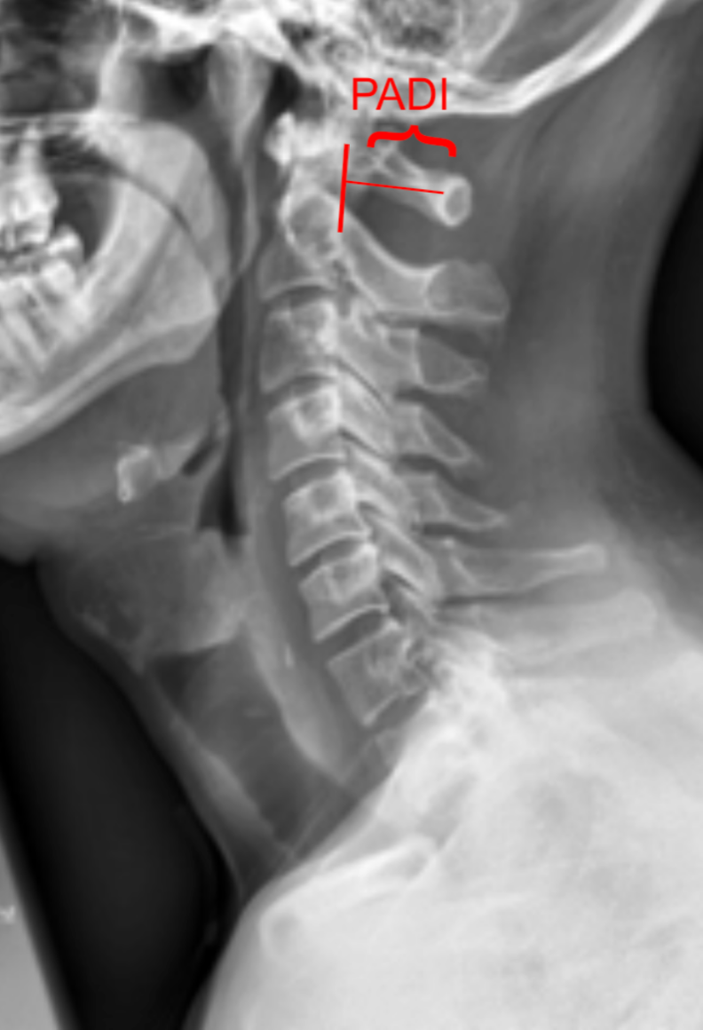
1) Description of Measurement
The Posterior Atlantodental Interval (PADI), also known as the space available for the cord (SAC), is the distance between the posterior surface of the odontoid process (dens) and the anterior surface of the posterior arch of the atlas (C1).
It represents the effective canal diameter at the atlantoaxial level and is a critical measurement for evaluating cervicomedullary compression. A decreased PADI suggests potential spinal cord impingement, even when the atlantodental interval (ADI) is within normal limits.
2) Instructions to Measure
Obtain a neutral lateral cervical spine X-ray (as shown in provided images).
Identify the posterior surface of the odontoid process (dens).
Identify the anterior surface of the posterior arch of C1 (atlas).
Draw a line along each of these two surfaces.
Measure the shortest perpendicular distance between them—this is the PADI.
For instability evaluation, repeat measurement on flexion and extension lateral views to assess for dynamic reduction in PADI.
3) Normal vs. Pathologic Ranges
Normal PADI (adults): ≥ 15 mm
Pathologic PADI (adults): < 14 mm, increased risk for spinal cord compression
Normal PADI (children): ≥ 13 mm
Pathologic PADI (children): < 13 mm, potential cord compression
4) Important References
Boden SD, Dodge LD, Bohlman HH, Rechtine GR. Rheumatoid arthritis of the cervical spine: a long-term analysis with predictors of paralysis and recovery. J Bone Joint Surg Am. 1993;75(9):1282–1297.
White AA, Panjabi MM. Clinical Biomechanics of the Spine. 2nd ed. Philadelphia, PA: Lippincott; 1990.
Dvorak J, Panjabi MM. Functional anatomy of the alar and transverse ligaments. Spine. 1987;12(2):183–189.
Smoker WRK. Craniovertebral junction: normal anatomy, craniometry, and congenital anomalies. Radiographics. 1994;14(2):255–277.
5) Other info....
PADI < 15 mm is associated with myelopathy and poor neurological outcomes in both traumatic and inflammatory (e.g. rheumatoid) atlantoaxial instability
Unlike the ADI, with reflects bony displacement, PADI more directly correlates with neurological compression
The PADI is a functional measure of canal space, unlike the ADI which measures ligamentous instability.
MRI correlation is strongly recommended when PADI is borderline (< 15 mm) to directly evaluate cord compression and signal changes.
In rheumatoid arthritis, progressive pannus formation can reduce PADI without major change in ADI, leading to late-onset myelopathy.
CT or MRI provides greater accuracy, but PADI on X-ray remains a simple and reproducible screening tool for instability and canal narrowing.
PADI < 10 mm almost always indicates neurologic compromise.