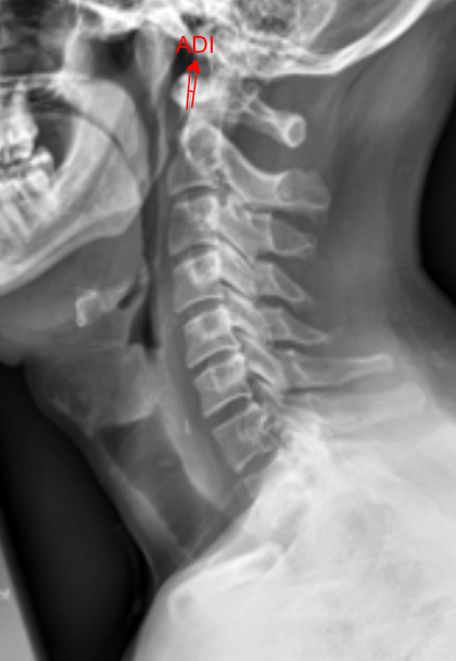
Image Type
Atlantodental Interval (ADI)
1) Description of Measurement
The Atlantodental Interval (ADI)—also known as the atlantoaxial interval (AAI)—is the distance between the posterior surface of the anterior arch of the atlas (C1) and the anterior surface of the odontoid process (dens) of the axis (C2).
This measurement assesses the integrity and stability of the transverse atlantal ligament (TAL), which stabilizes the atlantoaxial joint. An increased ADI suggests instability at the atlantoaxial junction, potentially due to trauma, rheumatoid arthritis, congenital anomalies, or ligamentous laxity.
2) Instructions to Measure
Obtain a neutral lateral cervical spine X-ray (as shown in the provided image).
Identify the anterior arch of C1 (atlas) and the odontoid process (dens) of C2.
Draw a line along the posterior border of the anterior arch of C1.
Draw a line along the anterior border of the odontoid process.
Measure the shortest distance between these two lines—this is the ADI.
Optional: Repeat the measurement on flexion and extension lateral views to evaluate dynamic instability.
3) Normal vs. Pathologic Ranges
Normal ADI (adults): ≤ 3 mm
Pathologic/Unstable ADI (adults): > 3 mm (suggests transverse ligament instability)
Normal ADI (children): ≤ 5 mm
Pathologic/Unstable ADI (children): > 5 mm (physiologic laxity greater than adults)
4) Important References
Fielding JW, Hawkins RJ. Atlanto-axial rotatory fixation (fixed rotatory subluxation of the atlanto-axial joint). J Bone Joint Surg Am. 1977;59(1):37–44.
White AA, Panjabi MM. Clinical Biomechanics of the Spine. 2nd ed. Philadelphia, PA: Lippincott; 1990.
Dvorak J, Panjabi MM. Functional anatomy of the alar and transverse ligaments. Spine. 1987;12(2):183–189.
Smoker WRK. Craniovertebral junction: normal anatomy, craniometry, and congenital anomalies. Radiographics. 1994;14(2):255–277.
5) Other info....
Dynamic (flexion-extension) change > 1 mm may also indicate instability, even if absolute values appear normal
Flexion-extension films are especially useful in detecting occult instability.
Increased ADI is commonly seen in trauma (transverse ligament rupture), rheumatoid arthritis, Down syndrome, or Grisel’s syndrome.
ADI is often accompanied by evaluation of the posterior atlantodental interval (PADI)—the distance between the posterior aspect of the dens and the anterior border of the posterior arch of C1.
PADI < 14 mm correlates with increased risk of spinal cord compression.
For preoperative planning, CT and MRI provide superior visualization of the odontoid, atlas ring, and ligamentous structures.