Image Type
Chamberlain’s Line / McGregor’s Line
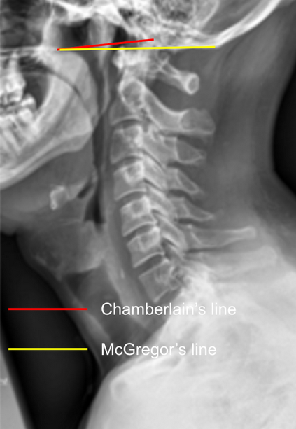
1) Description of Measurement
Chamberlain’s Line and McGregor’s Line are radiographic reference lines used to assess the position of the odontoid process (dens) relative to the skull base, helping identify basilar invagination or craniovertebral junction anomalies.
Chamberlain’s Line: Drawn from the posterior edge of the hard palate to the posterior margin of the foramen magnum (opisthion).
McGregor’s Line: Drawn from the posterior edge of the hard palate to the most caudal point of the occipital curve (inferior occipital surface).
The vertical distance of the dens tip above these lines indicates upward migration of the odontoid into the cranial cavity.
2) Instructions to Measure
Use a neutral lateral cervical spine X-ray (as shown in the provided image).
Identify landmarks:
Posterior hard palate (anterior reference)
Opisthion (posterior margin of foramen magnum) — for Chamberlain’s Line
Most caudal point of the occipital curve — for McGregor’s Line
Tip (apex) of the odontoid process (dens)
Draw the respective lines:
Chamberlain’s Line: From posterior hard palate → opisthion
McGregor’s Line: From posterior hard palate → most caudal occipital point
Measure the vertical distance (in mm) from the tip of the odontoid to each line:
Positive value = dens projects above the line
Negative value = dens lies below the line
3) Normal vs. Pathologic Ranges
Normal Chamberlain’s line: Dens ≤ 3 mm above line
Pathologic Chamberlain’s/Suggestive of basilar invagination: Dens > 3-5 mm above line
Normal McGregor’s line: Dens ≤ 4.5-5 mm above line
Pathologic McGregor’s/ Suggestive of basilar invagination: Dens > 4.5-5 mm above line
4) Important References
Chamberlain WE. Basilar impression (platybasia): a bizarre developmental anomaly of the occipital bone and upper cervical spine with remarks on the atlanto-occipital articulation. Yale J Biol Med. 1939;11:487–496.
McGregor M. The significance of certain measurements of the skull in the diagnosis of basilar impression. Br J Radiol. 1948;21:171–181.
Smoker WRK. Craniovertebral junction: normal anatomy, craniometry, and congenital anomalies. Radiographics. 1994;14(2):255–277.
Menezes AH. Craniovertebral junction anomalies: diagnosis and management. Semin Pediatr Neurol. 1997;4(3):209–223.
5) Other info....
Basilar invagination, atlantoaxial dislocation, or congenital anomalies (e.g., platybasia, Chiari malformation) should be considered when the dens projects above normal limits.
McGregor’s Line is more reproducible radiographically since the opisthion can be difficult to visualize on plain films.
Best assessed on neutral lateral X-rays or CT midsagittal reconstructions.
Always correlate with clinical findings (neurologic deficits, cervicomedullary compression) and adjunct imaging (MRI for soft tissue and neural structures).
Often measured alongside Wackenheim’s line (extension of the clivus) for comprehensive craniovertebral junction assessment.