Image Type
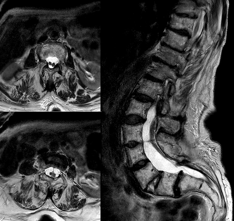
Diastematomyelia
1) Description of Measurement
Diastematomyelia, also termed split cord malformation, is a congenital form of spinal dysraphism in which the spinal cord is longitudinally divided into two hemicords over a variable segment. The split is caused by an intervening osseous, cartilaginous, or fibrous septum within the spinal canal. Each hemicord may have its own central canal, nerve roots, and vascular supply. The condition frequently results in cord tethering, progressive neurological dysfunction, and associated vertebral anomalies. The term split cord malformation is now preferred, as it includes both classic diastematomyelia and diplomyelia within the same spectrum.
2) Instructions to Measure
Perform MRI of the entire spine, with sagittal and axial T1- and T2-weighted sequences.
Identify:
Level(s) of cord splitting
Presence and type of intervening septum
Configuration of the dural sac(s)
Assess:
Whether the hemicords travel in one dural sac or two
Cranio-caudal extent of the split
Reunification of hemicords distally
Evaluate for associated findings:
Hydromyelia or syringomyelia
Low-lying conus and thickened filum
Spinal cord lipoma, dermoid, neurenteric cyst
Use CT to characterize bony spurs and vertebral anomalies.
Prenatal ultrasound may demonstrate a midline echogenic focus between posterior elements, suggesting a septum.
3) Normal vs. Pathologic Ranges
Normal
Single, continuous spinal cord
Single dural sac without midline septum
Pathologic
Longitudinal division of the cord into two hemicords
Classification:
Type I:
Two separate dural sacs
Rigid midline spur (osseous or cartilaginous)
Usually symptomatic
Type II:
Single dural sac
Nonrigid fibrous or fibrovascular septum
Often milder or asymptomatic
Typical anatomic distribution:
Most commonly L1–L3
Also seen at T7–T12
Conus medullaris lies below L2 in >75% of patients.
4) Important References
Pang D, Dias MS, Ahab-Barmada M. Split cord malformation: Part I: A unified theory of embryogenesis for double spinal cord malformations. Neurosurgery. 1992 Sep;31(3):451-80. doi: 10.1227/00006123-199209000-00010. PMID: 1407428.
Naidich TP, Harwood-Nash DC. Diastematomyelia: hemicord and meningeal sheaths; single and double arachnoid and dural tubes. AJNR Am J Neuroradiol. 1983 May-Jun;4(3):633-6. PMID: 6410818; PMCID: PMC8334888.
Adelson P. Diastematomyelia. Encyclopedia of the Neurological Sciences. 2014;:996-7. doi:10.1016/b978-0-12-385157-4.00741-7
Huang SL, He XJ, Wang KZ, Lan BS. Diastematomyelia: a 35-year experience. Spine (Phila Pa 1976). 2013 Mar 15;38(6):E344-9. doi: 10.1097/BRS.0b013e318283f6bc. PMID: 23492975.
Weston MJ. Spinal abnormalities. In: Twining P, McHugo JM, Pilling DW, editors. Textbook of fetal abnormalities. 2nd ed. Edinburgh: Churchill Livingstone; 2007. p. 143–173. doi:10.1016/B978-0-443-07416-5.50012-5.
5) Other info....
Embryologically linked to persistent accessory neuroenteric canal during weeks 3–4, explaining its strong association with vertebral anomalies like hemivertebrae, butterfly vertebrae, block vertebrae, and congenital scoliosis, often a presenting clue. It needs surgical correction before scoliosis surgery to prevent neurologic injury.