
Image Type
Syringomyelia
1) Description of Measurement
Syringomyelia is a disorder characterized by the formation of an abnormal, fluid-filled cavity (syrinx) within the central canal or parenchyma of the spinal cord. The syrinx develops due to disrupted cerebrospinal fluid (CSF) flow, most commonly at the craniocervical junction. Progressive expansion leads to compression and destruction of spinal cord pathways, classically beginning with decussating spinothalamic fibers. When the cavity extends into the brainstem, the condition is termed syringobulbia.
2) Instructions to Measure
Perform MRI using sagittal and axial T1- and T2-weighted sequences.
Identify the syrinx as a CSF-intensity cavity within the spinal cord.
Document:
Maximum anteroposterior diameter of the syrinx (mm)
Cranio-caudal extent (vertebral levels involved)
Location (central vs eccentric)
Presence of septations
Evaluate adjacent structures:
Posterior fossa and foramen magnum (Chiari malformation)
Spinal cord tumors or post-inflammatory adhesions
Use CT or radiographs as adjuncts to assess scoliosis or bony abnormalities.
3) Normal vs. Pathologic Ranges
Normal
No intramedullary fluid cavity
Central canal may be inapparent or minimally visible. Typically < 1–2 mm ("slit-like").
Pathologic
Any persistent intramedullary CSF-filled cavity. Generally defined as a cavity > 3 mm in width.
Larger diameter (>3–4 mm) and longer syrinx length correlate with higher symptom burden
Typical distribution: Most commonly between C2 and T9
Related entity:
Hydromyelia: dilation of the central canal that remains in communication with the fourth ventricle
Often associated with Chiari malformation (the most common underlying cause), tethered cord, or spinal tumors.
4) Important References
National Institute of Neurological Disorders and Stroke. Syringomyelia. Bethesda (MD): National Institute of Neurological Disorders and Stroke (US); [cited 2025 Dec 24]. Available from: https://www.ninds.nih.gov/health-information/disorders/syringomyelia
Fadila M, Sarrabia G, Shapira S, Yaacobi E, Baruch Y, Engel I, Ohana N. Orthopedic Manifestations of Syringomyelia: A Comprehensive Review. J Clin Med. 2025 May 1;14(9):3145. doi: 10.3390/jcm14093145. PMID: 40364175; PMCID: PMC12072671.
Klekamp J. How Should Syringomyelia be Defined and Diagnosed? World Neurosurg. 2018 Mar;111:e729-e745. doi: 10.1016/j.wneu.2017.12.156. Epub 2018 Jan 6. PMID: 29317358.
Shenoy VS, Munakomi S, Sampath R. Syringomyelia. [Updated 2024 Mar 14]. In: StatPearls [Internet]. Treasure Island (FL): StatPearls Publishing; 2025 Jan-. Available from: https://www.ncbi.nlm.nih.gov/books/NBK537110/
Greitz D. Unraveling the riddle of syringomyelia. Neurosurg Rev. 2006 Oct;29(4):251-63; discussion 264. doi: 10.1007/s10143-006-0029-5. Epub 2006 May 31. PMID: 16752160.
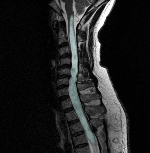
Figure 1. in: Shunting of recurrent post-traumatic syringomyelia into the fourth ventricle: a case report” by Chih-Lung Lin, Journal of Medical Case Reports, licensed under CC BY 2.0.