
Image Type
Hemivertebra Morphometry (wedge angle, segmentation)
1) Description of Measurement
Hemivertebra morphometry characterizes the degree of vertebral wedging and segmentation pattern of a hemivertebra, a congenital anomaly caused by failure of vertebral body formation. These parameters are critical in predicting curve progression, assessing deformity severity, and guiding timing and extent of surgical correction.
The measurement consists of both: Wedge angle — quantifying the angular deformity contributed by the hemivertebra and segmentation status — classifying the hemivertebra as fully segmented, semi-segmented, or unsegmented, based on the presence of adjacent disc spaces.
2) Instructions to Measure
A. Wedge Angle Measurement (Coronal MRI)
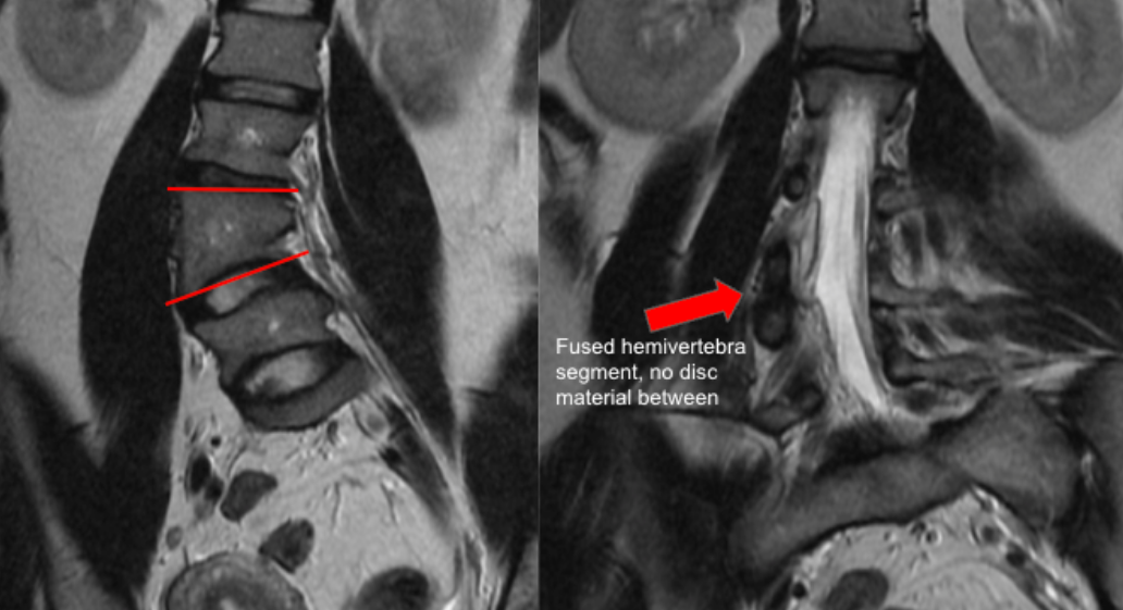
Select a coronal MRI slice that best visualizes the hemivertebra and adjacent vertebrae.
Identify the superior and inferior endplates of the hemivertebra.
Draw a line along the superior endplate of the hemivertebra.
Draw a second line along the inferior endplate of the hemivertebra.
Measure the acute angle between these two lines — this is the hemivertebral wedge angle (degrees).
B. Segmentation Assessment (Axial + Coronal MRI)
Evaluate the intervertebral discs above and below the hemivertebra.
Classify segmentation as:
Fully segmented: Disc spaces present both above and below
Semi-segmented: Disc present on only one side
Unsegmented (incarcerated): No disc spaces above or below, fused to adjacent vertebrae
3) Normal vs. Pathologic Ranges
Wedge Angle
Mild deformity: < 10°
Moderate deformity: 10° - 20°
Severe deformity / high progression risk: > 20°
4) Important References
Marks DS, Qaimkhani SA. The natural history of congenital scoliosis and kyphosis. Spine (Phila Pa 1976). 2009 Aug 1;34(17):1751-5. doi: 10.1097/BRS.0b013e3181af1caf.
Ruf M, Harms J. Hemivertebra resection by a posterior approach: innovative operative technique and first results. Spine (Phila Pa 1976). 2002 May 15;27(10):1116-23. doi: 10.1097/00007632-200205150-00020.
Basu PS, Elsebaie H, Noordeen MH. Congenital spinal deformity: a comprehensive assessment at presentation. Spine (Phila Pa 1976). 2002 Oct 15;27(20):2255-9. doi: 10.1097/00007632-200210150-00014.
5) Other info....
Fully segmented hemivertebrae with large wedge angles are most likely to cause progressive deformity.
Morphometry should be interpreted alongside:
Age of patient
Location (thoracic vs lumbar)
Presence of bars or rib anomalies
MRI is preferred for:
Evaluating associated spinal cord anomalies (tethered cord, syrinx, diastematomyelia)
These measurements inform decisions regarding:
Early hemivertebra excision
Growth-friendly instrumentation
Timing of fusion
Adapted from: Gaillard F, Fused hemivertebra. Case study, Radiopaedia.org (Accessed on 28 Dec 2025) https://doi.org/10.53347/rID-41487