
Image Type
Pavlov/Torg (Canal-Body Ratio)
1) Description of Measurement
The Pavlov–Torg ratio, also known as the canal–body ratio, is a dimensionless measurement used to assess cervical spinal canal stenosis by comparing the sagittal diameter of the spinal canal to the sagittal diameter of the corresponding vertebral body at the same level.
Originally described for lateral radiographs, this ratio can be accurately adapted to MRI, where it benefits from improved visualization of both bony and soft-tissue boundaries. The ratio minimizes errors from patient size and image magnification and serves as a screening tool for congenital or acquired cervical stenosis, which predisposes patients to cervical myelopathy and spinal cord injury.
2) Instructions to Measure
Select a true mid-sagittal cervical MRI slice (preferably T2-weighted) with clear visualization of:
Vertebral bodies
Posterior vertebral body margin / disc–osteophyte complex
Ligamentum flavum
Thecal sac
Choose the vertebral level of interest (typically C3–C7).
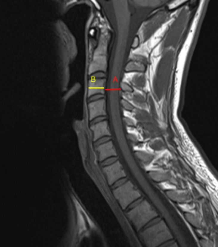
Measure the Sagittal Canal Diameter (A):
Measure the shortest anteroposterior distance between:
The posterior margin of the vertebral body or disc–osteophyte complex (anterior canal boundary), and
The anterior margin of the ligamentum flavum or lamina (posterior canal boundary).
Measure the Sagittal Vertebral Body Diameter (B):
Measure the anteroposterior diameter of the vertebral body at the same level, from the anterior cortex to the posterior cortex of the vertebral body.
Calculate the Pavlov–Torg Ratio:
Pavlov-Torg Ratio = AB
Repeat measurements at multiple levels (C3–C7) and record the lowest ratio, as this represents the level at greatest risk for stenosis.
3) Normal vs. Pathologic Ranges
Normal canal dimensions: ≥ 0.8
Borderline/relative stenosis: 0.7 - 0.9
Pathologic stenosis: < 0.7
Severe stenosis: ≤ 0.6; high myelopathy risk
Key points:
A ratio ≤ 0.7 is strongly associated with congenital cervical stenosis
MRI-based ratios may correlate better with cord compression and signal change than X-ray measurements
4) Important References
Pavlov H, Torg JS, Robie B, Jahre C. Cervical spinal stenosis: determination with vertebral body ratio method. Radiology. 1987 Sep;164(3):771-5. doi: 10.1148/radiology.164.3.3615879.
Lamothe G, Muller F, Vital JM, et al. Evolution of spinal cord injuries due to cervical canal stenosis without radiographic evidence of trauma (SCIWORET): a prospective study. Ann Phys Rehabil Med. 2011 Jun;54(4):213-24. English, French. doi: 10.1016/j.rehab.2011.02.003. Epub 2011 Mar 21.
Nouri A, Montejo J, Sun X, et al. Cervical Cord-Canal Mismatch: A New Method for Identifying Predisposition to Spinal Cord Injury. World Neurosurg. 2017 Dec;108:112-117. doi: 10.1016/j.wneu.2017.08.018. Epub 2017 Aug 12.
Boden SD, McCowin PR, Davis DO, et al. Abnormal magnetic-resonance scans of the cervical spine in asymptomatic subjects. A prospective investigation. J Bone Joint Surg Am. 1990 Sep;72(8):1178-84.
5) Other info....
The Pavlov–Torg ratio is a screening tool, not a definitive diagnostic measure
MRI provides superior assessment by incorporating:
Disc protrusions
Ligamentum flavum hypertrophy
True cord compression
A low ratio does not guarantee symptoms; many asymptomatic patients have ratios ≤ 0.7
Clinical correlation is essential and should include:
Neurologic exam
Cord signal change (T2 hyperintensity)
Absolute sagittal canal diameter
The ratio is especially useful in:
Athletes
Trauma risk stratification
Preoperative planning for cervical decompression
Adapted from: Di Muzio B, Normal cervical spine MRI. Case study, Radiopaedia.org (Accessed on 17 Dec 2025) https://doi.org/10.53347/rID-38418