
Image Type
Sagittal Canal Diameter
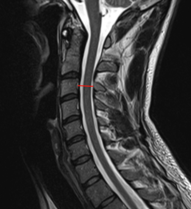
1) Description of Measurement
Sagittal canal diameter is a direct linear measurement of the anteroposterior (AP) dimension of the cervical spinal canal, reflecting the space available for the spinal cord. On MRI, this measurement incorporates both osseous and soft-tissue boundaries, making it the most accurate imaging modality for evaluating true canal compromise.
Reduced sagittal canal diameter correlates strongly with cervical spinal stenosis, myelopathy, and increased risk of neurologic injury, particularly in the presence of degenerative disc disease, posterior osteophytes, ligamentum flavum hypertrophy, or disc herniation.
2) Instructions to Measure
Select a mid-sagittal T2-weighted MRI slice that clearly visualizes:
Vertebral bodies
Posterior longitudinal ligament / disc margins
Ligamentum flavum
Thecal sac and spinal cord
Identify the vertebral level of interest (commonly C3–C7).
At the selected level:
Identify the posterior margin of the vertebral body or disc–osteophyte complex (anterior canal boundary).
Identify the anterior margin of the ligamentum flavum or lamina (posterior canal boundary).
Using digital calipers, measure the shortest anteroposterior distance (mm) between these two structures.
Repeat measurements at multiple levels if needed and record the smallest diameter, as this represents the most clinically relevant stenotic level.
3) Normal vs. Pathologic Ranges
Normal Canal: >13 mm
Relative/moderate stenosis: 10-12 mm
Absolute stenosis: <12 mm
Severe stenosis: < 8 mm; high risk for myelopathy
4) Important References
Hinck VC, Sachdev NS. Developmental stenosis of the cervical spinal canal. Brain. 1966 Mar;89(1):27-36. doi: 10.1093/brain/89.1.27.
Pavlov H, Torg JS, Robie B, Jahre C. Cervical spinal stenosis: determination with vertebral body ratio method. Radiology. 1987 Sep;164(3):771-5. doi: 10.1148/radiology.164.3.3615879.
Takao T, Morishita Y, Okada S, et al. Clinical relationship between cervical spinal canal stenosis and traumatic cervical spinal cord injury without major fracture or dislocation. Eur Spine J. 2013 Oct;22(10):2228-31. doi: 10.1007/s00586-013-2865-7. Epub 2013 Jun 23.
Boden SD, McCowin PR, Davis DO, et al. Abnormal magnetic-resonance scans of the cervical spine in asymptomatic subjects. A prospective investigation. J Bone Joint Surg Am. 1990 Sep;72(8):1178-84.
5) Other info....
MRI is the gold standard for assessing canal diameter due to visualization of:
Disc herniations
Ligamentum flavum hypertrophy
Cord compression and intramedullary signal change
Should be interpreted alongside:
Spinal cord signal (T2 hyperintensity)
Pavlov/Torg ratio (X-ray screening tool)
Clinical signs of myelopathy
Dynamic factors (motion-dependent narrowing) may not be fully captured on static MRI—consider flexion-extension X-rays when instability is suspected
Diameter alone does not dictate symptoms; cord deformation and chronicity are critical modifiers
Adapted from: Di Muzio B, Normal cervical spine MRI. Case study, Radiopaedia.org (Accessed on 17 Dec 2025) https://doi.org/10.53347/rID-38418