Image Type
C2-C7 Sagittal Vertical Axis (cSVA) (cervical contribution)
1) Description of Measurement
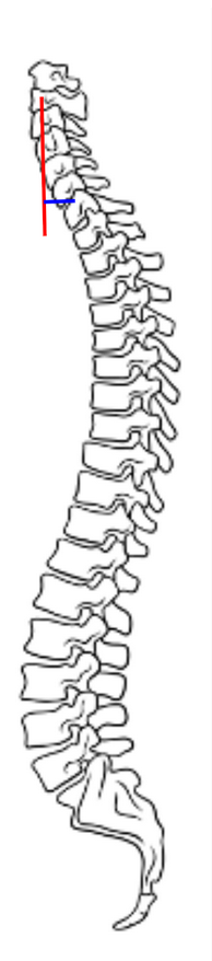
The C2–C7 Sagittal Vertical Axis (cSVA) quantifies the sagittal alignment of the cervical spine and represents the horizontal distance between the C2 plumb line and the C7 vertebral body on a standing lateral full-spine X-ray.
It provides insight into cervical sagittal balance and how the cervical spine contributes to overall sagittal alignment.
An increased cSVA indicates anterior translation of the head relative to the cervical spine, reflecting positive sagittal malalignment, often associated with forward head posture, cervical deformity, or compensatory changes to maintain horizontal gaze.
This measurement complements global sagittal balance parameters such as C7–S1 SVA and thoracic kyphosis and is critical for understanding regional-to-global alignment relationships.
2) Instructions to Measure
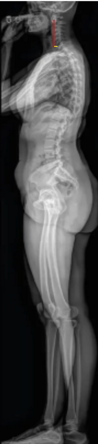
Obtain a standing lateral full-spine X-ray, ensuring the patient’s gaze is horizontal, knees are extended, and arms are supported forward to prevent superimposition.
Identify key landmarks:
C2 centroid: the midpoint of the C2 vertebral body, typically midway between the anterior and posterior cortices.
C7 posterior–superior corner: the most posterior–superior point of the C7 vertebral body.
Drop a vertical plumb line (parallel to the gravitational vertical) from the C2 centroid.
Measure the horizontal distance (in millimeters) between this vertical line and the posterior–superior corner of C7.
If the C2 plumb line lies anterior to C7, record as a positive cSVA (forward head posture).
If the plumb line lies posterior, record as negative cSVA (posterior translation, rare).
Document both the direction and magnitude of deviation.
Optional: For global assessment, correlate C2–C7 SVA with T1 slope, C2–C7 Cobb angle, and C7–S1 SVA to determine compensatory cervical alignment relative to thoracolumbar posture.
3) Normal vs. Pathologic Ranges
Normal cSVA: 10-20 mm; balanced cervical alignment; neutral head position
Mild positive imbalance: > 20-40 mm; mild anterior head translation; possible compensatory lordosis
Pathologic/positive imbalance: > 40 mm; significant anterior shift; associated with cervical deformity and worse outcomes
Negative cSVA: < 0 mm; posterior translation; may occur after overcorrection or hyperlordosis
Key Point:
A cSVA > 40 mm is associated with increased Neck Disability Index (NDI) scores and clinical symptoms of sagittal imbalance (Tang et al., 2012).
4) Important References
Tang JA, Scheer JK, Smith JS, et al. The impact of standing regional cervical sagittal alignment on outcomes in posterior cervical fusion surgery. Neurosurgery. 2012;71(3):662–669.
Ames CP, Blondel B, Scheer JK, et al. Cervical radiographical alignment: comprehensive assessment techniques and potential importance in cervical myelopathy. Spine. 2013;38(22 Suppl 1):S149–160.
Smith JS, Shaffrey CI, Ames CP, et al. Assessment of cervical sagittal alignment and clinical implications. J Neurosurg Spine. 2013;19(2):141–159.
Iyer S, Lenke LG, Nemani VM, et al. Impact of cervical sagittal alignment parameters on patient-reported outcomes after posterior cervical fusion. Spine. 2016;41(23):1790–1798.
Kato M, Namikawa T, Matsumura A, et al. C2–C7 sagittal vertical axis and cervical tilt: relationships with cervical alignment and clinical outcomes. Spine. 2016;41(3):E160–E168.
5) Other info....
C2–C7 SVA isolates the cervical spine’s contribution to overall sagittal alignment, distinct from the global C7–S1 SVA.
Increased cSVA correlates with worse functional outcomes and greater muscular fatigue from maintaining horizontal gaze.
C2–C7 SVA strongly correlates with T1 slope and C2–C7 lordosis; greater T1 slope often necessitates greater lordosis to maintain balance.
Restoration of physiological cSVA is a major goal in cervical deformity correction surgery.
Postoperatively, large positive cSVA is linked to loss of horizontal gaze, increased pain, and compensatory thoracic extension.
Consistent measurement technique (neutral posture, defined landmarks) is essential for accurate follow-up comparison.
Advanced modalities like EOS imaging and 3D reconstruction can further refine accuracy and assess coupled cervical-thoracolumbar relationships.