Image Type
Coronal Balance
1) Description of Measurement
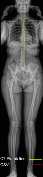
Coronal Balance is a radiographic parameter used to assess lateral (side-to-side) alignment of the spine in the coronal plane. It quantifies how well the head and trunk are centered over the pelvis when viewed from the front (AP or PA full-length spine X-ray).
It is an essential measurement for evaluating scoliosis, compensatory curves, and postoperative deformity correction.
Coronal balance helps determine whether the spine is globally aligned (balanced) or deviated laterally (imbalanced), reflecting the relationship between the C7 vertebral body and the central sacral vertical line (CSVL).
2) Instructions to Measure
Obtain a standing full-length AP or PA spine X-ray showing the entire spine, pelvis, and femoral heads (as shown in the provided image).
Identify the following landmarks:
C7 vertebral body center: mark the midpoint of the C7 vertebral body.
Center of the sacrum (S1 endplate): mark the midpoint of the superior sacral endplate.
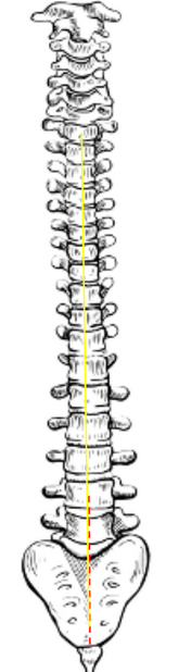
Draw the Central Sacral Vertical Line (CSVL): a vertical plumb line extending upward from the center of the sacrum (S1).
Draw a vertical line downward from the center of the C7 vertebral body (the C7 plumb line).
Measure the horizontal distance (in millimeters) between the C7 plumb line and the CSVL — this is the Coronal Balance.
A C7 plumb line that passes through or near the CSVL indicates coronal balance.
A plumb line deviated to the right or left indicates coronal imbalance.
Note the direction of deviation (right or left).
3) Normal vs. Pathologic Ranges
Normal coronal balance: ≤ 20 mm (2 cm); head and trunk are centered over pelvis, physiologic alignment
Coronal imbalance: > 20 mm (2 cm); significant lateral deviation; compensatory or structural deformity
Note direction (right or left imbalance); indicates deviation direction from the CSVL
4) Important References
Glassman SD, Berven S, Bridwell K, Horton W, Dimar JR. Coronal and sagittal balance in idiopathic scoliosis: does correction correlate with improvement in pain and function? Spine. 2005;30(7):682–688.
Schwab FJ, Lafage V, Boyce R, Skalli W, Farcy JP. Gravity line analysis in adult volunteers: age-related correlation with spinal parameters, pelvic parameters, and foot position. Spine. 2006;31(25):E959–E967.
Hresko MT, Talwalkar VR, Schwend RM. Clinical practice guideline: evaluation and treatment of idiopathic scoliosis in children and adolescents. J Am Acad Orthop Surg. 2014;22(9):577–587.
Lafage V, Schwab F, Skalli W, Hawkinson N, Gagey PM, Ondra S. Standing balance and alignment in adult spinal deformity: analysis of spinopelvic and gravity line parameters. Spine. 2008;33(14):1572–1578.
5) Other info....
Coronal Balance provides a global assessment of spinal alignment, complementing sagittal balance measures such as Sagittal Vertical Axis (SVA).
In scoliosis, the goal of correction is to restore the C7 plumb line to within 2 cm of the CSVL, indicating balanced correction.
Pelvic obliquity or leg length discrepancy can affect coronal balance — these should be corrected or accounted for when measuring.
Postoperative coronal imbalance can lead to compensatory trunk shift, shoulder asymmetry, or impaired functional outcomes.
EOS low-dose imaging or stitched long-cassette radiographs are preferred for accurate, full-body coronal assessment.
Always record the direction and magnitude of imbalance, as well as whether the deviation is structural (curve rigidity) or compensatory (pelvic or leg-length related).