
Image Type
Pedicle Dimensions
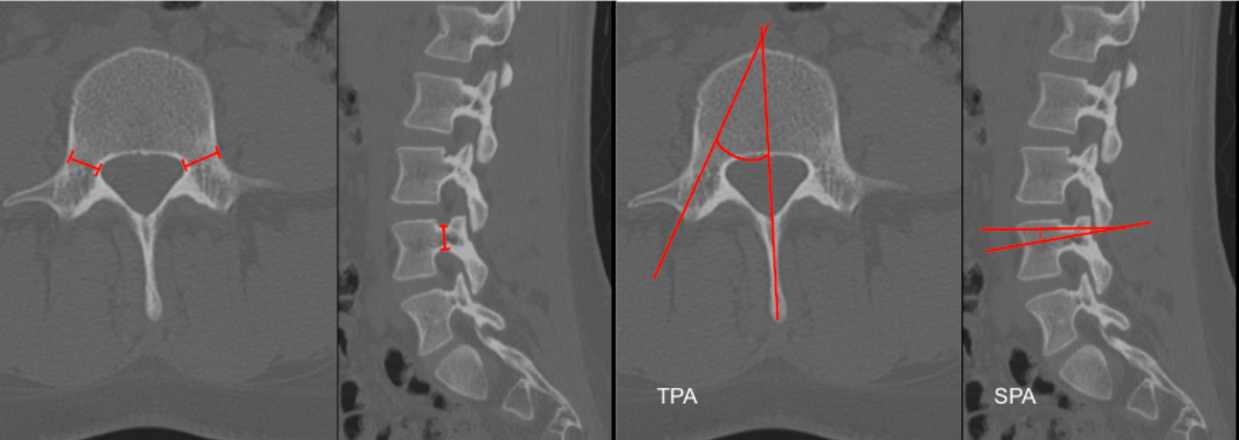
1) Description of Measurement
Lumbar pedicle morphometry quantifies the size and orientation of the pedicles and is essential for safe pedicle screw placement. Measurements include:
Pedicle width (transverse diameter)
Pedicle height (sagittal diameter)
Transverse pedicle angle (TPA) – determines medial–lateral screw trajectory
Sagittal pedicle angle (SPA) – determines cephalocaudal screw trajectory
2) Instructions to Measure
A. Pedicle Width (Transverse Diameter) – Axial CT
Select the axial slice at the pedicle mid-body level.
Identify the medial and lateral cortices of the pedicle.
Measure the minimum transverse distance (mm) between cortices.
B. Pedicle Height (Sagittal Diameter) – Sagittal CT
On parasagittal CT, identify the pedicle.
Measure the minimum superior–inferior distance (mm) between pedicle cortices.
C. Transverse Pedicle Angle – Axial CT
Draw a line through the pedicle axis.
Draw a reference line along the posterior vertebral body wall.
Measure the angle between these lines — this is the medial angulation angle.
D. Sagittal Pedicle Angle – Sagittal CT
Draw a line along the pedicle axis.
Draw a horizontal reference line through the posterior vertebral body wall.
Measure the angle — this is the cephalocaudal trajectory angle.
3) Normal vs. Pathologic Ranges
L1: Width (6-8 mm); Height (10-12 mm); TPA (5-10°); SPA (0-5°)
L2: Width (7-9 mm); Height (11-13 mm); TPA (10-15°); SPA (0-5°)
L3: Width (8-11 mm); Height (12-14 mm); TPA (15-20°); SPA (5-10°)
L4: Width (9-13 mm); Height (13-15 mm); TPA (20-30°); SPA (5-10°)
L5: Width (12-18 mm); Height (14-16 mm); TPA (25-35°); SPA (10-15°)
Key point:
Pedicle width < 5 mm significantly increases risk of cortical breach.
4) Important References
Zindrick MR, Wiltse LL, Doornik A, et al. Analysis of the morphometric characteristics of the thoracic and lumbar pedicles. Spine (Phila Pa 1976). 1987 Mar;12(2):160-6. doi: 10.1097/00007632-198703000-00012.
Sugisaki K, An HS, Espinoza Orías AA, et al. In vivo three-dimensional morphometric analysis of the lumbar pedicle isthmus. Spine (Phila Pa 1976). 2009 Nov 15;34(24):2599-604. doi: 10.1097/BRS.0b013e3181b52a37.
Coscia A, Paige K, Hostetter M, et al. Transverse Pedicle Angle Is Associated With Pelvic Incidence and Increased in Lumbar Isthmic Spondylolisthesis. Global Spine J. 2022 Apr;12(3):359-365. doi: 10.1177/2192568220951190.
Liu L, Wang H, Wang Q, et al. A Study of the Sagittal Angle of Lumbar Bicortical Pedicle Screws from the Anatomic Perspective of the Lumbar Artery. World Neurosurg. 2019 May;125:e435-e441. doi: 10.1016/j.wneu.2019.01.100.
5) Other info....
Always measure each pedicle individually — asymmetry is common in deformity and degenerative disease.
Use the smallest pedicle width to select screw diameter (typically 80% of pedicle width).
Adapted from: Feger J, Campos A, Murphy A, et al. CT lumbar spine (protocol). Reference article, Radiopaedia.org (Accessed on 03 Jan 2026) https://doi.org/10.53347/rID-90041