
Image Type
Cross-Sectional Area of Canal (CSA)
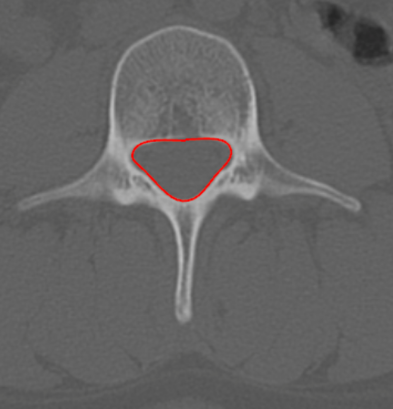
1) Description of Measurement
Lumbar spinal canal CSA on CT quantifies the bony cross-sectional area available for the thecal sac and cauda equina. It is a robust morphologic parameter for diagnosing lumbar spinal stenosis, particularly useful when MRI is contraindicated or to characterize osseous encroachment from facet hypertrophy, laminar thickening, or congenital canal narrowing.
2) Instructions to Measure
Identify the lumbar level of greatest canal narrowing on sagittal reconstructions.
Navigate to the corresponding axial CT slice at the mid-vertebral body or disc level.
Using a freehand or polygon ROI tool, trace the inner cortical margin of the bony spinal canal, including the laminae, pedicles, and posterior vertebral body margin.
Close the ROI to compute the cross-sectional area (mm²).
Record the smallest CSA across all lumbar levels.
3) Normal vs. Pathologic Ranges
Normal CSA: > 120 mm2
Borderline narrowing: 100 - 120 mm2
Lumbar spine stenosis: < 100 mm2
Severe stenosis: < 75 mm2
Critical stenosis: < 50 mm2
4) Important References
Maeder B, Becce F, Kehtari S, et al. Evolution of the Cross-Sectional Area of the Osseous Lumbar Spinal Canal across Decades: A CT Study with Reference Ranges in a Swiss Population. Diagnostics (Basel). 2023 Feb 15;13(4):734. doi: 10.3390/diagnostics13040734.
Cho J, Kang KN, Lee MS, Kim YU. Surgical versus nonsurgical management of lumbar degenerative spondylolisthesis based on spinal canal cross-sectional area. Medicine (Baltimore). 2024 Jan 12;103(2):e36874. doi: 10.1097/MD.0000000000036874.
Genevay S, Atlas SJ. Lumbar spinal stenosis. Best Pract Res Clin Rheumatol. 2010 Apr;24(2):253-65. doi: 10.1016/j.berh.2009.11.001.
5) Other info....
CT-based CSA primarily reflects bony stenosis; it does not account for ligamentum flavum or disc material — correlate with MRI dural sac CSA for neural compromise.
Should be interpreted in conjunction with:
AP canal diameter
Lateral recess width
Foraminal dimensions
Adapted from: Feger J, Campos A, Murphy A, et al. CT lumbar spine (protocol). Reference article, Radiopaedia.org (Accessed on 03 Jan 2026) https://doi.org/10.53347/rID-90041