
Image Type
Filum Terminale Thickness
1) Description of Measurement
The filum terminale is a thin fibrous structure extending from the conus medullaris to the coccyx, anchoring the spinal cord. Abnormal thickening of the filum terminale is a key imaging feature of tethered cord syndrome (TCS) and may cause progressive neurologic deterioration due to traction on the spinal cord.
2) Instructions to Measure
Identify the conus medullaris tip on mid-sagittal images.
Follow the filum terminale inferiorly to the upper sacral canal (typically S1–S2 level).
On the slice where the filum is best visualized, use an electronic caliper to measure the maximum transverse diameter of the filum terminale.
Record the thickness in millimeters (mm).
3) Normal vs. Pathologic Ranges
Normal thickness: ≤ 2 mm
Suggestive of TCS: > 2 mm
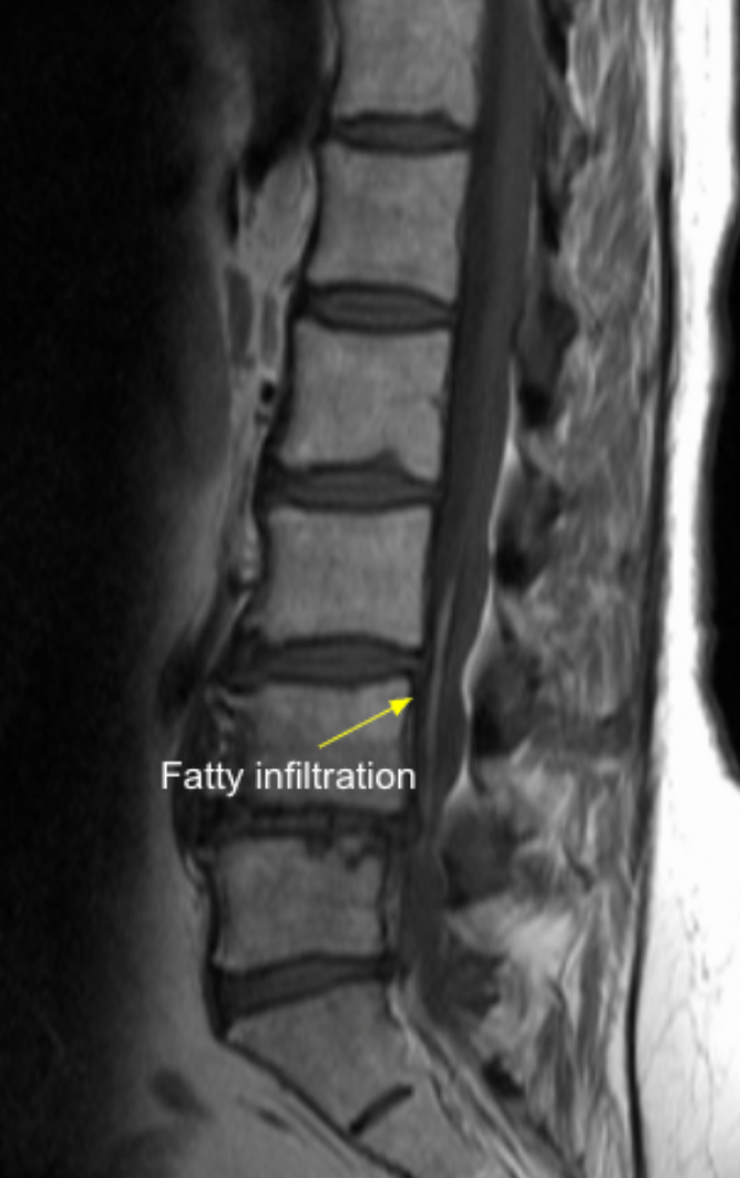
Highly suspicious for TCS: Fatty infiltration present
4) Important References
Yamada S, Won DJ, Siddiqi J, Yamada SM. Tethered cord syndrome: overview of diagnosis and treatment. Neurol Res. 2004 Oct;26(7):719-21. doi: 10.1179/016164104225017947.
Hertzler DA 2nd, DePowell JJ, Stevenson CB, Mangano FT. Tethered cord syndrome: a review of the literature from embryology to adult presentation. Neurosurg Focus. 2010 Jul;29(1):E1. doi: 10.3171/2010.3.FOCUS1079.
Grimme JD, Castillo M. Congenital anomalies of the spine. Neuroimaging Clin N Am. 2007 Feb;17(1):1-16. doi: 10.1016/j.nic.2006.11.002.
5) Other info....
Filum thickness should always be interpreted alongside:
Conus medullaris level
Neurologic symptoms
Associated dysraphic anomalies (lipoma, dermal sinus)
A thickened filum with a low-lying conus is highly diagnostic of tethered cord syndrome.
Adapted from: Kecler-Pietrzyk A, Lipoma of filum terminale. Case study, Radiopaedia.org (Accessed on 03 Jan 2026) https://doi.org/10.53347/rID-53131