
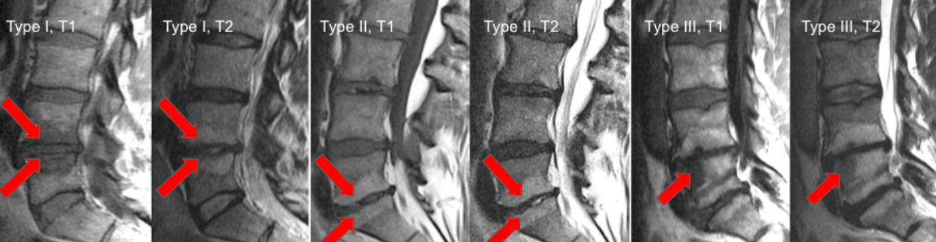
Image Type
Modic Endplate Changes (Type I, II, III)
1) Description of Measurement
Modic changes describe signal intensity alterations of vertebral body marrow adjacent to degenerated intervertebral discs, reflecting different stages of the degenerative cascade. They are strongly associated with disc degeneration, low-back pain, and segmental instability.
Three distinct patterns are recognized based on MRI signal characteristics.
2) Instructions to Measure
Identify vertebral endplates adjacent to symptomatic or degenerated discs.
Evaluate marrow signal on:
T1-weighted images
T2-weighted images
Fat-suppressed or STIR sequences
Classify the signal pattern at each affected endplate:
Type I: T1 hypointense, T2 hyperintense; edema / active inflammation
Type II: T1 hyperintense, T2 iso-/hyperintense; fatty marrow replacement
Type III: T1 hypointense, T2 hypointense; subchondral sclerosis
Record the Modic type, level, and laterality.
3) Normal vs. Pathologic Ranges
No signal change: normal endplate
Type I: acute degenerative/inflammatory change
Type II: Chronic degenerative change
Type III: Advanced degenerative change with sclerosis
Key points:
Type I often correlates with active pain generators.
Type III is least common and reflects end-stage disease.
4) Important References
Modic MT, Steinberg PM, Ross JS, et al. Degenerative disk disease: assessment of changes in vertebral body marrow with MR imaging. Radiology. 1988 Jan;166(1 Pt 1):193-9. doi: 10.1148/radiology.166.1.3336678.
Jensen TS, Karppinen J, Sorensen JS, et al. Vertebral endplate signal changes (Modic change): a systematic literature review of prevalence and association with non-specific low back pain. Eur Spine J. 2008 Nov;17(11):1407-22. doi: 10.1007/s00586-008-0770-2.
Hopayian K, Raslan E, Soliman S. The association of modic changes and chronic low back pain: A systematic review. J Orthop. 2022 Nov 17;35:99-106. doi: 10.1016/j.jor.2022.11.003.
5) Other info....
Modic changes often co-localize with Pfirrmann grade IV–V discs and reduced Disc-Height Index.
Type I lesions may respond differently to targeted therapies (e.g., anti-inflammatory strategies).
Report Modic changes in conjunction with:
Disc herniation size
Segmental alignment
Dural sac CSA
Adapted from: Dudli S, Fields AJ, Samartzis D, et al. Pathobiology of Modic changes. Eur Spine J. 2016 Nov;25(11):3723-3734. doi: 10.1007/s00586-016-4459-7.