
Image Type
Disc-Height Index (DHI)
1) Description of Measurement
The Disc-Height Index (DHI) is a normalized metric that quantifies intervertebral disc height relative to adjacent vertebral body height. By expressing disc height as a ratio, DHI minimizes magnification and patient-size variability and serves as a sensitive marker for disc degeneration, collapse, and segmental instability.
2) Instructions to Measure
Identify the disc level to be evaluated on the mid-sagittal MRI.
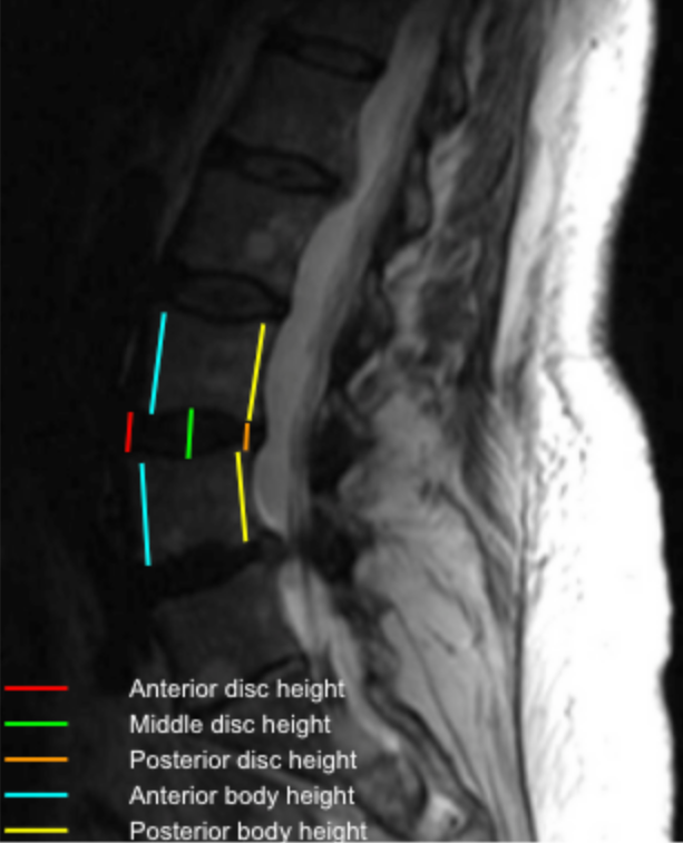
Measure disc height at three locations:
Anterior disc height (A)
Middle disc height (M)
Posterior disc height (P)
Calculate the mean disc height (DH):
DH = (A + M + P)/3
Measure the heights of the vertebral bodies immediately above (V₁) and below (V₂) the disc at their midpoints.
Calculate the Disc-Height Index (DHI):
DHI = DH/(V1 + V2)/2
3) Normal vs. Pathologic Ranges
Normal DHI: ≥ 0.40
Mild disc height loss: 0.30 - 0.39
Moderate degeneration: 0.20 - 0.29
Severe disc collapse: < 0.20
Key points:
Progressive reduction in DHI correlates with Pfirrmann grades IV–V, facet arthropathy, and instability.
Comparison to adjacent segments improves accuracy.
4) Important References
Chen X, Sima S, Sandhu HS, et al. Radiographic evaluation of lumbar intervertebral disc height index: An intra and inter-rater agreement and reliability study. J Clin Neurosci. 2022 Sep;103:153-162. doi: 10.1016/j.jocn.2022.07.018.
Snowden R, Miller J, Saidon T, et al. Does index level sagittal alignment determine adjacent level disc height loss? J Neurosurg Spine. 2019 Jun 21;31(4):579-586. doi: 10.3171/2019.4.SPINE181468.
Chen IR, Wei TS. Disc height and lumbar index as independent predictors of degenerative spondylolisthesis in middle-aged women with low back pain. Spine (Phila Pa 1976). 2009 Jun 1;34(13):1402-9. doi: 10.1097/BRS.0b013e31817b8fbd.
5) Other info....
DHI is useful for longitudinal studies to track disc degeneration progression.
Should be reported together with:
Pfirrmann grade
Modic endplate changes
Segmental alignment (spondylolisthesis, kyphosis)
Adapted from: Feger J, Er A, Yap J, et al. Lumbar spine protocol (MRI). Reference article, Radiopaedia.org (Accessed on 01 Jan 2026) https://doi.org/10.53347/rID-147093