
Image Type
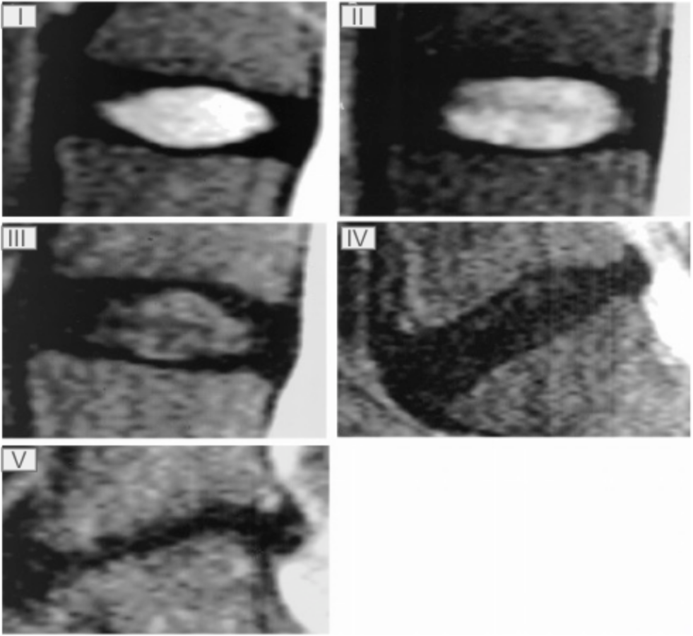
Pfirrmann Classification (I-V)
1) Description of Measurement
The Pfirrmann Classification is a five-grade MRI-based system that characterizes lumbar intervertebral disc degeneration using disc structure, signal intensity on T2-weighted images, distinction between nucleus pulposus and annulus fibrosus, and disc height. It provides a standardized, reproducible method to quantify disc degeneration and correlate imaging findings with clinical symptoms and biomechanical deterioration
2) Instructions to Measure
Review the mid-sagittal and parasagittal T2-weighted MRI sequences of the lumbar spine.
For each disc level (L1–2 through L5–S1), assess:
Overall disc structure (homogeneous vs inhomogeneous)
Signal intensity of the nucleus pulposus
Distinction between nucleus and annulus
Disc height compared with adjacent normal levels
Assign a grade according to the following criteria:
Grade I: Homogeneous bright white nucleus; clear nucleus–annulus distinction, T2 hyperintense, similar to CSF; normal disc height
Grade II: Inhomogeneous ± horizontal bands in disc; clear nucleus-annulus distinction; T2 hyperintense disc; normal disc height
Grade III: Inhomogenous, gray; Unclear nucleus-annulus distinction; intermediate T2 signal; normal/slightly decreased disc height
Grade IV: Inhomogenous, gray-black; no nucleus-annulus distinction; intermediate to hypointense T2 signal; normal/moderately decreased disc height
Grade V: Inhomogenous, black disc; no nucleus-annulus distinction; T2 hypointense signal; collapsed disc height
3) Normal vs. Pathologic Ranges
Grade I-II: Normal/minimal degeneration
Grade III: Early degenerative disc disease
Grade IV: Advanced disc degeneration
Grade V: Severe disc degeneration with collapse
Key points:
Grades IV-V are strongly associated with biomechanical instability, modic endplate changes, and higher likelihood of symptomatic disease
4) Important References
Pfirrmann CW, Metzdorf A, Zanetti M, et al. Magnetic resonance classification of lumbar intervertebral disc degeneration. Spine (Phila Pa 1976). 2001 Sep 1;26(17):1873-8. doi: 10.1097/00007632-200109010-00011.
Miyazaki M, Hong SW, Yoon SH, et al. Reliability of a magnetic resonance imaging-based grading system for cervical intervertebral disc degeneration. J Spinal Disord Tech. 2008 Jun;21(4):288-92. doi: 10.1097/BSD.0b013e31813c0e59.
Griffith JF, Wang YX, Antonio GE, et al. Modified Pfirrmann grading system for lumbar intervertebral disc degeneration. Spine (Phila Pa 1976). 2007 Nov 15;32(24):E708-12. doi: 10.1097/BRS.0b013e31815a59a0.
5) Other info....
Grading is performed on T2-weighted mid-sagittal images, as disc hydration best reflects degeneration.
The Pfirrmann system demonstrates excellent inter- and intraobserver reliability (κ ≈ 0.69–0.90).
In equivocal cases, integrate disc height, nucleus signal, and structure together rather than relying on a single feature.
Adapted from: Pfirrmann CW, Metzdorf A, Zanetti M, et al. Magnetic resonance classification of lumbar intervertebral disc degeneration. Spine (Phila Pa 1976). 2001 Sep 1;26(17):1873-8. doi: 10.1097/00007632-200109010-00011.