
Image Type
Foraminal Height and Width
1) Description of Measurement
Lumbar foraminal height and width quantify the available space for the exiting nerve root within the neural foramen. Reduction in either dimension results in foraminal stenosis, most commonly due to disc height loss, facet hypertrophy, osteophytes, or spondylolisthesis, and is a major cause of radicular leg pain.
These measurements allow objective grading of foraminal stenosis as mild, moderate, or severe.
2) Instructions to Measure
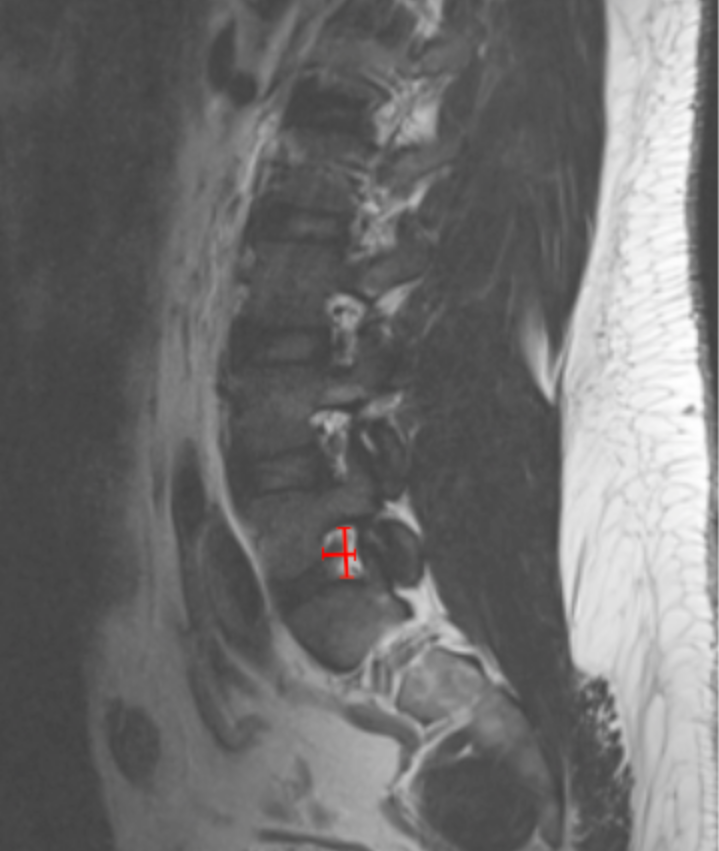
On sagittal T2-weighted MRI, identify the disc level corresponding to the patient’s radicular symptoms.
Select the parasagittal slice that best visualizes the neural foramen.
Measure Foraminal Height:
Identify the inferior cortex of the pedicle above and the superior cortex of the pedicle below.
Measure the vertical distance (mm) between these two points.
Measure Foraminal Width:
On the same slice, identify the posterior vertebral body/disc margin (anterior boundary) and the anterior aspect of the superior articular facet (posterior boundary).
Measure the anteroposterior distance (mm).
Corroborate with axial T2 images to assess root compression or obliteration of perineural fat.
3) Normal vs. Pathologic Ranges
Foraminal Height
Normal: ≥ 15 mm
Mild Stenosis: 12-15 mm
Moderate Stenosis: 8-12 mm
Severe Stenosis: < 8 mm
Foraminal Width
Normal: ≥ 4 mm
Mild Stenosis: 3-4 mm
Moderate Stenosis: 2-3 mm
Severe Stenosis: < 2 mm
Key points:
Severe stenosis is typically associated with loss of perineural fat and direct nerve root compression.
Height reduction reflects disc collapse, whereas width reduction reflects facet and osteophyte encroachment.
4) Important References
Lee S, Lee JW, Yeom JS, et al. A practical MRI grading system for lumbar foraminal stenosis. AJR Am J Roentgenol. 2010 Apr;194(4):1095-8. doi: 10.2214/AJR.09.2772.
Seo J, Lee JW. Magnetic Resonance Imaging Grading Systems for Central Canal and Neural Foraminal Stenoses of the Lumbar and Cervical Spines With a Focus on the Lee Grading System. Korean J Radiol. 2023 Mar;24(3):224-234. doi: 10.3348/kjr.2022.0351.
Park HJ, Kim SS, Han CH, Lee SY, Chung EC, Kim MS, Kwon HJ. The clinical correlation of a new practical MRI method for grading cervical neural foraminal stenosis based on oblique sagittal images. AJR Am J Roentgenol. 2014 Aug;203(2):412-7. doi: 10.2214/AJR.13.11647.
5) Other info....
Foraminal stenosis is best assessed on parasagittal MRI.
Should be evaluated together with:
Lateral recess width
Dural sac CSA
Dynamic foraminal collapse may be underestimated in supine MRI; consider weight-bearing or extension MRI in equivocal cases.
Adapted from: Feger J, Er A, Yap J, et al. Lumbar spine protocol (MRI). Reference article, Radiopaedia.org (Accessed on 01 Jan 2026) https://doi.org/10.53347/rID-147093