
Image Type
Lateral Recess Width/Depth
1) Description of Measurement
Lateral recess width (also termed lateral recess depth) quantifies the available space for the traversing nerve root between the posterior vertebral body/disc complex and the superior articular facet. This is the most common site of nerve root compression in degenerative lumbar spinal stenosis and is particularly sensitive to facet hypertrophy, ligamentum flavum thickening, and disc bulge.
This measurement directly reflects the risk of radiculopathy and complements central canal CSA in evaluating symptomatic stenosis.
2) Instructions to Measure
Use the sagittal MRI to identify the lumbar level where symptoms or stenosis are most pronounced.
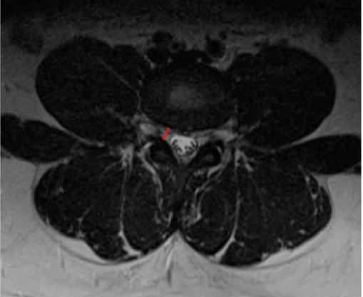
Scroll to the corresponding axial T2-weighted slice at the level of the pedicle and disc.
Identify:
The medial border of the superior articular facet (posterior boundary).
The posterolateral margin of the vertebral body or disc (anterior boundary).
Draw a straight line between these two points, oriented along the expected path of the traversing nerve root.
Measure the distance in millimeters (mm) — this is the lateral recess width/depth.
3) Normal vs. Pathologic Ranges
Normal: ≥ 5 mm
Relative Stenosis: 3 - 5 mm
Severe lateral recess stenosis: < 3 mm
Critical stenosis: Visible root impingement/obliteration
Key points:
Values < 3 mm strongly correlate with symptomatic radiculopathy.
The side with the smallest width should be documented.
4) Important References
Spinnato P, Petrera MR, Parmeggiani A, et al. A new comprehensive MRI classification and grading system for lumbosacral central and lateral stenosis: clinical application and comparison with previous systems. Radiol Med. 2024 Jan;129(1):93-106. doi: 10.1007/s11547-023-01741-3.
Caprariu R, Oprea MD, Poenaru DV, Andrei D. Correlation between Preoperative MRI Parameters and Oswestry Disability Index in Patients with Lumbar Spinal Stenosis: A Retrospective Study. Medicina (Kaunas). 2023 Nov 14;59(11):2000. doi: 10.3390/medicina59112000.
Goni VG, Hampannavar A, Gopinathan NR, et al. Comparison of the oswestry disability index and magnetic resonance imaging findings in lumbar canal stenosis: an observational study. Asian Spine J. 2014 Feb;8(1):44-50. doi: 10.4184/asj.2014.8.1.44.
5) Other info....
Lateral recess stenosis is often the primary driver of unilateral leg symptoms.
Should be evaluated with:
Dural sac CSA
Foraminal area
Central canal diameter may appear preserved even when lateral recess width is critically narrowed; therefore this measurement is essential in symptomatic patients.
Adapted from: Feger J, Er A, Yap J, et al. Lumbar spine protocol (MRI). Reference article, Radiopaedia.org (Accessed on 01 Jan 2026) https://doi.org/10.53347/rID-147093