
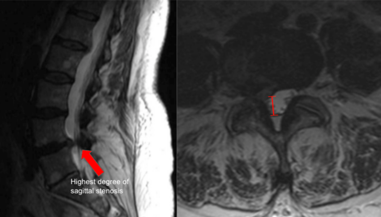
Image Type
Anteroposterior (AP) Canal Diameter
1) Description of Measurement
Lumbar AP canal diameter quantifies the anteroposterior dimension of the central spinal canal and is a standard morphologic measurement for diagnosing lumbar spinal stenosis. It reflects the available space for the cauda equina and is reduced by disc bulge, facet hypertrophy, ligamentum flavum thickening, or spondylolisthesis.
This measurement is most reliable on axial T2-weighted MRI, where cerebrospinal fluid provides natural contrast.
2) Instructions to Measure
Use the sagittal MRI to identify the disc level of maximal stenosis.
Scroll to the corresponding axial T2 slice at that level.
Identify:
The posterior margin of the vertebral body or disc (anterior boundary).
The anterior margin of the ligamentum flavum or lamina (posterior boundary).
Draw a straight line between these two points along the midline of the canal.
Measure this distance in millimeters (mm) — this is the AP canal diameter.
3) Normal vs. Pathologic Ranges
Normal: ≥ 12 mm
Relative (moderate) stenosis: 10 - 12 mm
Absolute (severe) stenosis: < 10 mm
Critical stenosis: < 7 mm
Key points:
Symptoms typically correlate when diameter is < 10 mm.
AP diameter underestimates severity when compression is asymmetric; consider canal area as adjunct.
4) Important References
Abel F, Tan ET, Chazen JL, et al. MRI after Lumbar Spine Decompression and Fusion Surgery: Technical Considerations, Expected Findings, and Complications. Radiology. 2023 Jul;308(1):e222732. doi: 10.1148/radiol.222732.
Hansen BB, Nordberg CL, Hansen P, et al. Weight-bearing MRI of the Lumbar Spine: Spinal Stenosis and Spondylolisthesis. Semin Musculoskelet Radiol. 2019 Dec;23(6):621-633. doi: 10.1055/s-0039-1697937.
Genevay S, Atlas SJ. Lumbar spinal stenosis. Best Pract Res Clin Rheumatol. 2010 Apr;24(2):253-65. doi: 10.1016/j.berh.2009.11.001.
5) Other info....
Measurement should be interpreted alongside:
Cross-sectional canal area
Lateral recess diameter
Foraminal dimensions
Central canal stenosis is dynamic and may worsen with extension.
Report:
Level of worst stenosis
Laterality of compression
Associated findings (facet hypertrophy, disc extrusion, spondylolisthesis)
Adapted from: Feger J, Er A, Yap J, et al. Lumbar spine protocol (MRI). Reference article, Radiopaedia.org (Accessed on 01 Jan 2026) https://doi.org/10.53347/rID-147093